Click the image below for a high-res guide to pediatric brain tumors, particularly tailored to those medical students studying for the USMLE Step 1 exam.
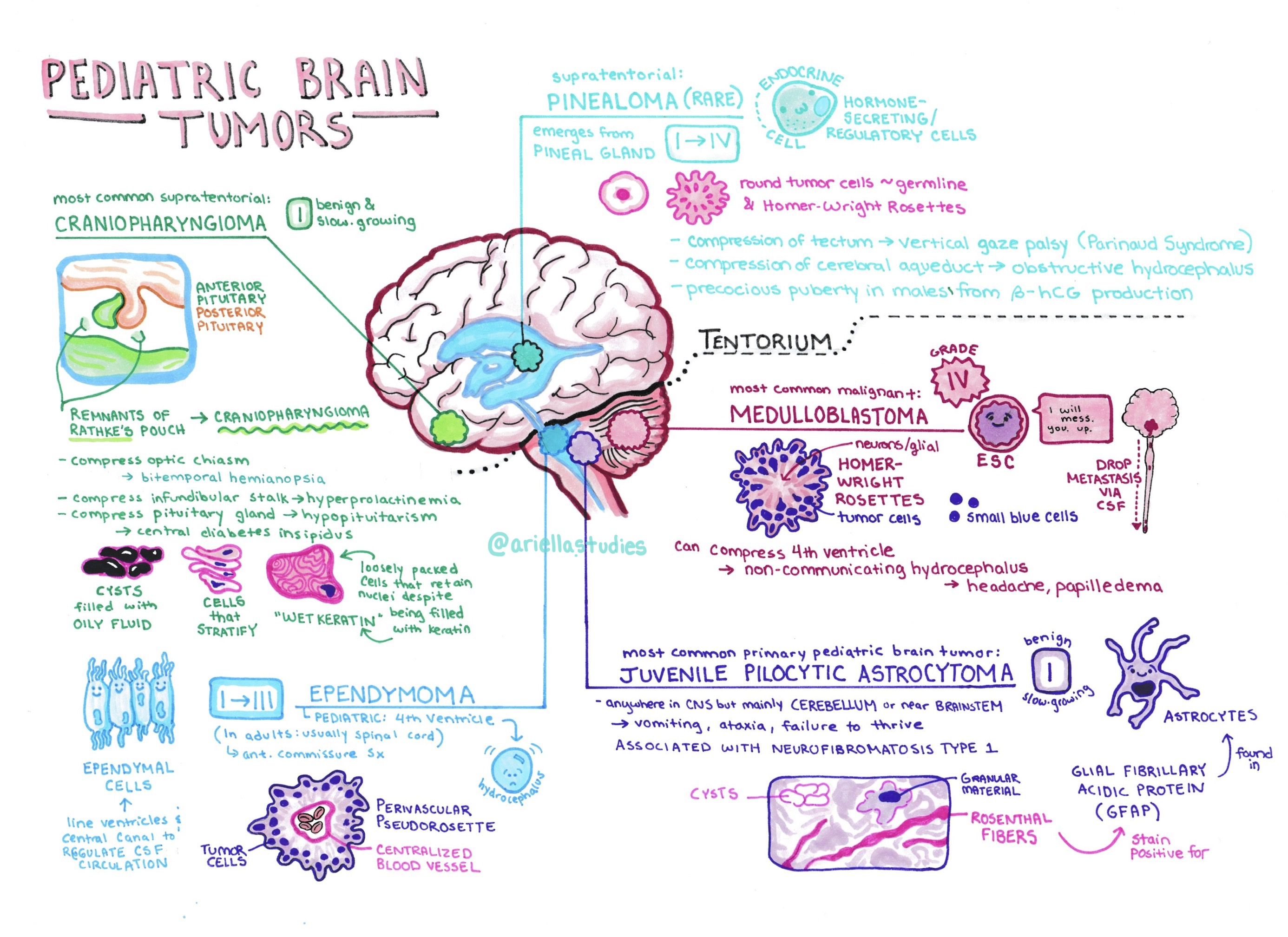
Describes: Craniopharyngioma and pinealoma above the tentorium; ependymoma, medulloblastoma, and juvenile pill cystic astrocytoma below. Locations are mapped on an anatomical illustration of the brain in sagittal cross-section. Cell types, grades, and other attributes are noted. Histological highlights are reproduced semi-accurately.
References: primarily the OsmosisMed video on pediatric brain tumors, supplemented with Amboss notes and classroom learning at WUSM. Special thanks to Osmosis for making the cells in their videos so adorable that I was inspired to try to emulate their style. I recommend watching the Osmosis video and then using this guide to condense and review the material.
Materials: Staedtler Triplus fineliners, Copic Markers.
Photographed and touched up in Adobe Photoshop.